Summary
This project aimed to develop an AI system capable of analyzing medical images, such as X-rays, MRIs, and CT scans, to detect and diagnose various types of cancer. The system was designed to assist radiologists and medical professionals in identifying cancerous lesions with high accuracy and efficiency. By leveraging advanced convolutional neural networks (CNN) and transfer learning, the AI system sought to enhance diagnostic capabilities, reduce human error, and streamline the workflow in medical imaging departments.
The Challenge
Cancer diagnosis through medical imaging is a complex and critical task that demands high accuracy and expertise. Radiologists face significant challenges due to the sheer volume of images to analyze, the subtlety of early-stage cancer lesions, and the need for rapid, precise diagnosis. The primary challenges included:
1. Volume and Variety of Data: Managing and processing large datasets of medical images from different modalities and sources.
2. Annotation and Validation: Ensuring accurate annotation of images by medical experts, which is time-consuming and labor-intensive.
3. Model Performance: Developing a robust AI model that can generalize well across various types of cancer and imaging conditions.
4. Explainability and Trust: Making the AI’s decision-making process transparent and trustworthy for medical professionals.
5. Integration and Compliance: Seamlessly integrating the AI system into existing hospital infrastructure while ensuring compliance with healthcare regulations.
The Solution
The solution was a comprehensive AI system designed with a well-structured workflow to address each challenge effectively:
1. Data Collection:
– Gathered a diverse dataset of medical images, encompassing different cancer types and stages, from multiple reputable sources.
2. Data Annotation:
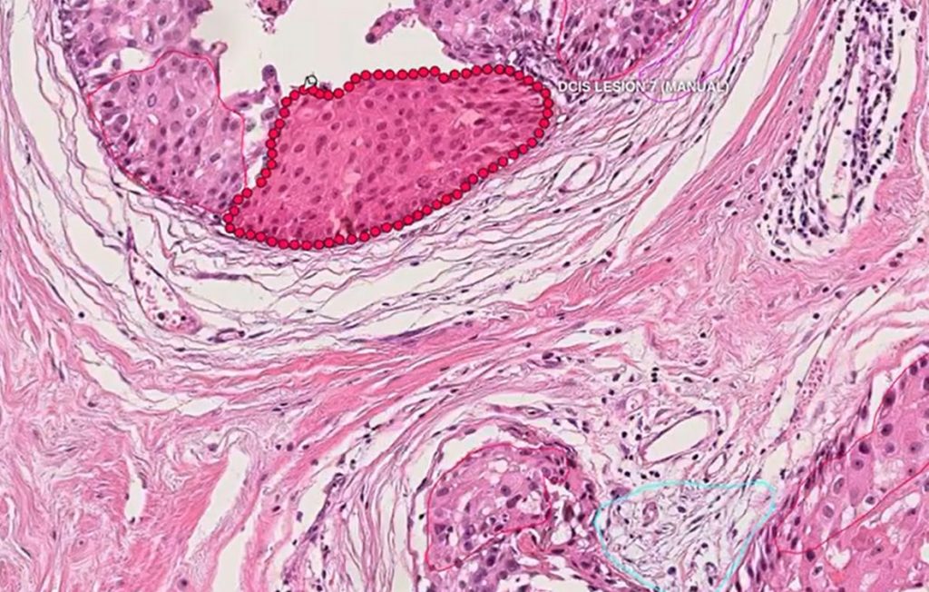
– Collaborated with medical experts to annotate images meticulously, marking cancerous regions and adding essential metadata.
3. Model Development:
- Chose appropriate CNN architectures such as ResNet and Inception.
- Applied transfer learning techniques using pre-trained models to leverage existing knowledge.
- Trained the model on the annotated dataset, incorporating data augmentation techniques to improve robustness and handle variations in the data.
4. Model Evaluation:
- Split the dataset into training, validation, and test sets to ensure comprehensive evaluation.
- Employed cross-validation and performance metrics (e.g., accuracy, sensitivity, specificity) to assess the model.
- Fine-tuned the model based on validation feedback to enhance performance.
5. Explainability and Validation:
- Implemented Grad-CAM to visualize the model’s decision-making process, providing insights into how the AI identifies cancerous lesions.
- Validated the model’s predictions with medical professionals to ensure clinical relevance and accuracy.
6. Deployment:
- Developed a user-friendly web-based interface for easy access by medical staff.
- Integrated the AI system with existing hospital infrastructure using APIs.
- Ensured data security and compliance with healthcare regulations (e.g., HIPAA, GDPR).
The Outcome
The AI system significantly improved the efficiency and accuracy of cancer detection and diagnosis in medical imaging. Key outcomes included:
1. Enhanced Diagnostic Accuracy:
– The AI system achieved high accuracy rates in detecting various types of cancer, reducing false positives and false negatives.
2. Increased Efficiency:
– Automated analysis of medical images streamlined the workflow, allowing radiologists to focus on more complex cases and improving overall productivity.
3. Improved Clinical Decision-Making:
– Grad-CAM visualizations and explainable AI techniques provided radiologists with valuable insights into the AI’s decision-making process, fostering trust and enabling better-informed clinical decisions.
4. Seamless Integration:
– The web-based interface and API integration ensured that the AI system could be easily adopted by hospitals, enhancing existing diagnostic processes without significant disruptions.
5. Regulatory Compliance:
– The system adhered to stringent data security and privacy regulations, ensuring patient data was protected and compliant with healthcare standards.
In conclusion, the AI system for analyzing medical images to detect cancer successfully addressed the critical challenges in medical imaging, offering a powerful tool for early and accurate cancer diagnosis. The project demonstrated the potential of AI in transforming healthcare by enhancing diagnostic capabilities, improving efficiency, and supporting medical professionals in delivering better patient outcomes.