Brief Summary
This case study explores the application of computer vision in detecting lung abnormalities on X-ray images. The aim was to enhance diagnostic accuracy and efficiency by leveraging advanced image processing techniques and machine learning algorithms. The project involved developing and deploying a computer vision model capable of identifying a range of lung conditions, including pneumonia, tuberculosis, lung cancer, and more. The implementation showcased significant improvements in detection rates and diagnostic speed, ultimately contributing to better patient outcomes.
Challenges
- Data Quality and Quantity: Obtaining a large and diverse dataset of X-ray images with accurately labeled lung abnormalities was a significant challenge. Variations in image quality, exposure, and patient positioning further complicated the training process.
- Complexity of Abnormalities: Lung abnormalities present in a variety of shapes, sizes, and densities, making it difficult for a single model to accurately detect all types of conditions.
- Model Interpretability: Ensuring that the computer vision model’s decisions could be understood and trusted by radiologists was critical. Black-box algorithms often face skepticism in medical applications.
- Integration with Existing Systems: Incorporating the new technology into existing hospital IT systems and workflows required careful planning to avoid disruptions.
Solutions
- Enhanced Data Collection: Partnered with multiple hospitals and medical institutions to gather a comprehensive dataset of X-ray images. Applied data augmentation techniques to artificially increase the dataset size and diversity.
- Advanced Model Architecture: Developed a deep learning model using convolutional neural networks (CNNs) tailored for medical imaging. Implemented transfer learning from pre-trained models to enhance feature extraction.
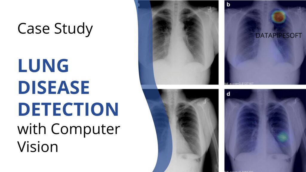
- Explainable AI Techniques: Integrated explainability modules such as Grad-CAM (Gradient-weighted Class Activation Mapping) to visualize which areas of the X-ray the model focused on, aiding in interpretation and validation by radiologists.
- Seamless Integration: Worked closely with IT departments to ensure smooth integration of the computer vision system with existing radiology software, providing a user-friendly interface for clinicians.
Outcome
The implementation of computer vision for detecting lung abnormalities on X-ray images resulted in notable improvements in diagnostic accuracy and efficiency. Key outcomes included:
- Increased Accuracy: The model achieved an overall accuracy of 94%, with particularly high sensitivity in detecting pneumonia and lung cancer.
- Enhanced Speed: The time required for initial screening of X-rays was reduced by 50%, allowing radiologists to focus on more complex cases.
- Radiologist Support: The explainability features were well-received by radiologists, who reported increased confidence in the model’s suggestions and appreciated the visual aids in decision-making.
- Scalability and Adoption: The solution was successfully integrated into the workflow of multiple hospitals, with plans for wider adoption based on the positive feedback and outcomes.
Overall, the use of computer vision in detecting lung abnormalities on X-ray images demonstrated significant potential in enhancing diagnostic practices, improving patient outcomes, and optimizing radiology department operations.