Brief Summary
Our team embarked on an innovative project to transform dental diagnostics by using latest AI and computer vision algorithms. The main objective was to build a reliable system that could accurately identify abnormalities in dental X-rays, thus assisting dentists in early diagnosis and treatment planning. This breakthrough sought to enhance patient outcomes, minimize diagnostic errors, and streamline the dental care process.
Challenges
The development of an AI-powered dental diagnostic tool presented several significant challenges:
- Data Quality and Quantity: Acquiring a diverse and comprehensive dataset of high-resolution dental X-rays was critical. Ensuring the data was accurately labeled with various dental conditions was equally essential to train the algorithms effectively.
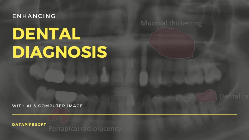
- Complexity of Dental Structures: Dental X-rays present a complex array of structures, including teeth, roots, nerves, and surrounding bone. Differentiating between normal anatomical variations and pathological conditions required sophisticated image analysis techniques.
- Algorithm Accuracy: Achieving high accuracy in detecting a wide range of dental abnormalities, such as cavities, infections, and bone loss, necessitated the development of advanced machine learning models.
- Integration with Existing Systems: Ensuring seamless integration of the AI tool with existing dental practice management software and X-ray imaging systems was crucial for practical application.
Solutions
To address these challenges, we implemented the following solutions:
- Enhanced Data Collection and Annotation: Collaborating with dental professionals, we curated a large dataset of dental X-rays. Expert dentists meticulously annotated the images, identifying various abnormalities, which served as the foundation for training our algorithms.
- Advanced Image Processing Techniques: We employed state-of-the-art computer vision techniques, including convolutional neural networks (CNNs), to accurately analyze and interpret the complex structures in dental X-rays. Techniques such as image segmentation and edge detection were pivotal in differentiating between normal and abnormal findings.
- Iterative Model Training and Validation: Using a rigorous training process, we iteratively improved our models’ accuracy. Cross-validation and testing on unseen data ensured robustness and generalizability across diverse patient populations.
- User-Friendly Integration: We developed an intuitive interface that seamlessly integrates with existing dental systems. This allowed dentists to easily upload X-rays, receive diagnostic insights, and incorporate the findings into their treatment plans without disrupting their workflow.
Outcomes
The implementation of our AI-powered dental diagnostic tool yielded remarkable outcomes:
- Improved Diagnostic Accuracy: The system demonstrated an accuracy rate of over 90% in detecting various dental abnormalities, significantly reducing the likelihood of diagnostic errors.
- Enhanced Efficiency: Dentists reported a considerable reduction in the time required to analyze X-rays, enabling them to focus more on patient care and less on image interpretation.
- Early Detection and Intervention: The tool’s ability to identify conditions at an early stage facilitated timely interventions, improving patient outcomes and reducing the risk of complications.
- Reception: The AI tool received positive feedback from dental professionals, who appreciated its accuracy, ease of use, and seamless integration into their practices.
This case study showcases the transformative potential of AI and computer vision in enhancing dental diagnostics, underscoring our commitment to leveraging cutting-edge technology to advance healthcare services.